Leg Anatomy Muscles Ligaments And Tendons / Ankle Pain - One of the most important tendons is the quadriceps tendon.. The gastrocnemius is the larger calf muscle, forming the bulge visible beneath the skin. A joint capsule is a watertight sac that surrounds a joint. Originates from the lateral condyle of the tibia and the medial surface of the fibula. Skeletal muscles are held to the bones with the help of tendons. There are many muscles located in the lower leg, but there are three that are particularly well known—the gastrocnemius and the soleus, which are the most powerful muscles in the lower leg, and the anterior tibialis.
One of the most important tendons is the quadriceps tendon. The major tendons include the following: This lies on the front of the knee and connects the quadriceps muscles of the thigh to the tibia via the patella and patellar ligament (or tendon). There are four muscles in the anterior compartment of the leg. The foot is a part of vertebrate anatomy which serves the purpose of supporting the animal's weight and allowing for locomotion on land.
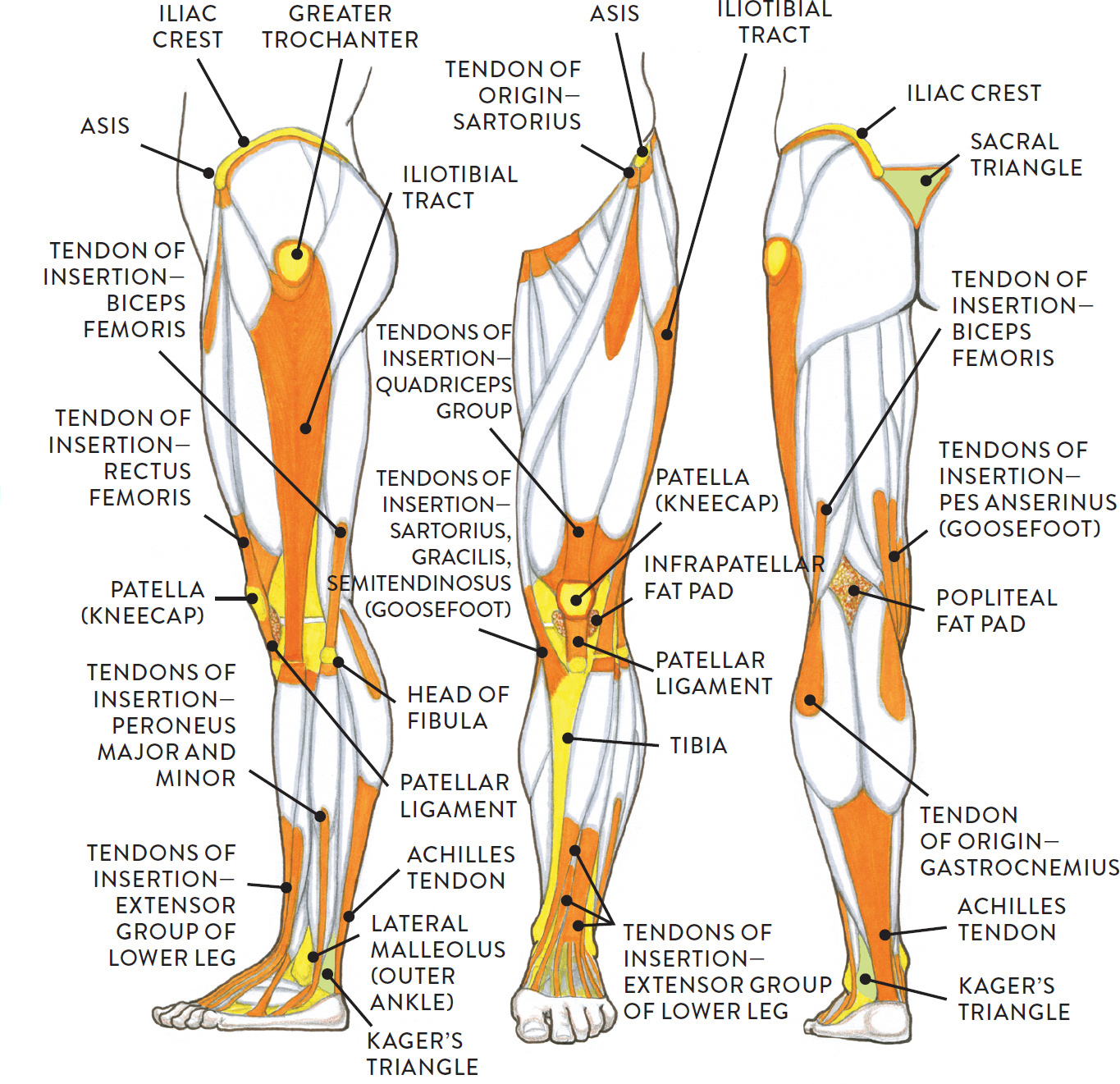
Upper and lower leg, three views from schoolbag.info In humans, the foot is one of the most complex structures in the body. The calcaneofibular ligament (cfl), which connects the calcaneus, or heel bone, to the fibula. This important tendon in the back of the calf and ankle connects the plantaris, gastrocnemius, and soleus muscles to. Possibly the most important tendon in terms of mobility is the achilles tendon. Ligaments, muscles and tendons keep us connected and help us move. One of the most important tendons is the quadriceps tendon. Tendons attach muscle to bone. The tarsal bones are found near the.
This lies on the front of the knee and connects the quadriceps muscles of the thigh to the tibia via the patella and patellar ligament (or tendon).
The calcaneofibular ligament (cfl), which connects the calcaneus, or heel bone, to the fibula. The popliteofibular ligament attaches the popliteus tendon to the fibular head and has a thickness similar to the lateral collateral ligament (fig. Related posts of muscle, tendons and ligaments of leg human muscle gross anatomy. Skeletal muscles are held to the bones with the help of tendons. Muscles, either individually or in groups, are supported by fascia. There are four major ligaments that surround the knee joint. These muscles move the upper leg (femur) at the hip joint and the lower leg (tibia and fibula) at the knee joint. Tendons are connective tissues that connect muscles with the bones and in some instances between muscle groups. In the hip, the joint capsule is formed by a group of three strong ligaments that connect the femoral head to the acetabulum. The calf muscles, through the achilles tendon, are the main plantarflexors of the ankle which pulls the foot down. The quadriceps and hamstring muscles work together to straighten (extend) and bend (flex) the leg. It provides the power necessary to straighten the knee. Leg anatomy muscles ligaments and tendons :
The adductor muscles pull the legs together. Ligaments, tendons, and muscles play an important role in the function of the hip. The foot is a part of vertebrate anatomy which serves the purpose of supporting the animal's weight and allowing for locomotion on land. The last of the muscle compartments of the lower leg is the lateral compartment (figure 15) is comprised of two muscles, the peroneus longus and the peroneus brevis. In humans, the foot is one of the most complex structures in the body.
Tendons and Ligaments of Ox Leg | ClipArt ETC from etc.usf.edu Skeletal muscles are attached to the bones by tendons. This lies on the front of the knee and connects the quadriceps muscles of the thigh to the tibia via the patella and patellar ligament (or tendon). Tendons attach muscle to bone. Skeletal muscles are held to the bones with the help of tendons. The soft tissue in the knee joint (tendons, ligaments, menisci, cartilage) that provides stability in the knee and hold the bones. Tendons vary in size and are somewhat elastic and attach bones to muscles. In the leg, muscle strains happen when a muscle is either stretched beyond its limits or forced into extreme contraction. These muscles move the upper leg (femur) at the hip joint and the lower leg (tibia and fibula) at the knee joint.
Tendons and ligaments are bands of connective tissue that help stabilize the body and allow movement.
Tendons and ligaments are bands of connective tissue that help stabilize the body and allow movement. Possibly the most important tendon in terms of mobility is the achilles tendon. Flexion, extension, medial rotation, and lateral rotation) and it connects the tibia and the fibula, with the thigh bone (femur). The lower leg lies between the knee and the ankle. There are four major ligaments that surround the knee joint. The gastrocnemius is the larger calf muscle, forming the bulge visible beneath the skin. It is a pivotal hinge joint in the leg that allows for a variety of movements (i.e. Ligaments are structures that connect two bones together. The calf muscle, on the back of the lower leg, is actually made up of two muscles: The tarsal bones are found near the. The foot is a part of vertebrate anatomy which serves the purpose of supporting the animal's weight and allowing for locomotion on land. Kegel muscle anatomy 12 photos of the kegel muscle anatomy kegel muscle anatomy, human muscles, kegel muscle anatomy. Skeletal muscles are attached to the bones by tendons.
Because the leg has many different muscles, it is vulnerable to several different types of muscle strains. Tendons attach muscle to bone. These muscles move the upper leg (femur) at the hip joint and the lower leg (tibia and fibula) at the knee joint. Because the leg has many different muscles, it is vulnerable to several different types of muscle strains. To better understand foot and leg muscle/tendon injuries, it is important to appreciate the basic elements that enable your body parts to move.
Instant Anatomy - Lower Limb - Areas/Organs - Lower Leg ... from www.instantanatomy.net This lies on the front of the knee and connects the quadriceps muscles of the thigh to the tibia via the patella and patellar ligament (or tendon). The quadriceps and hamstring muscles work together to straighten (extend) and bend (flex) the leg. Related posts of muscles and tendons of the leg kegel muscle anatomy. The popliteofibular ligament attaches the popliteus tendon to the fibular head and has a thickness similar to the lateral collateral ligament (fig. Thigh muscle strain anatomy the thigh has three sets of strong muscles: Ligaments are soft tissue structures that connect bones to bones. To better understand foot and leg muscle/tendon injuries, it is important to appreciate the basic elements that enable your body parts to move. Because the leg has many different muscles, it is vulnerable to several different types of muscle strains.
You hear them referred to as your gams, poles or limbs. but, whatever you call them, your legs are composed of bones, muscles, tendons and ligaments.
Thigh muscle strain anatomy the thigh has three sets of strong muscles: The leg anatomy includes the quads, hams, glutes, hip flexors, adductors & abductors. Ligaments connect two or more bones together and help stabilize joints. Related posts of muscles and tendons of the leg kegel muscle anatomy. There are four major ligaments that surround the knee joint. The calf muscles (gastrocnemius and soleus), which are connected to the calcaneus via the achilles tendon. Kegel muscle anatomy 12 photos of the kegel muscle anatomy kegel muscle anatomy, human muscles, kegel muscle anatomy. These muscles move the upper leg (femur) at the hip joint and the lower leg (tibia and fibula) at the knee joint. Achilles tendon, which attaches the calf muscle and calcaneus. Deltoid ligaments, which attach the tibia to the talus and calcaneus and provide stability to the insides of the ankles. This lies on the front of the knee and connects the quadriceps muscles of the thigh to the tibia via the patella and patellar ligament (or tendon). Leg anatomy muscles ligaments and tendons : The hamstring muscles in the back of the thigh, the quadriceps muscles in the front, and the adductor (groin) muscles on the inside.