Home » Without Label » Pelvic Anatomy : Anatomy: Pelvic Floor Muscles | Bekkenbodem, Lichaam ... - The superior two thirds correspond to the uterine body and the inferior third to the cervix.
Pelvic Anatomy : Anatomy: Pelvic Floor Muscles | Bekkenbodem, Lichaam ... - The superior two thirds correspond to the uterine body and the inferior third to the cervix.
Pelvic Anatomy : Anatomy: Pelvic Floor Muscles | Bekkenbodem, Lichaam ... - The superior two thirds correspond to the uterine body and the inferior third to the cervix.. This mri male pelvis axial cross sectional anatomy tool is absolutely free to use. If you continue browsing the site, you agree to the use of cookies on this website. • pelvis begins at the iliac crests and ends at the symphysis pubis. Use the mouse scroll wheel to move the images up and down alternatively use the tiny arrows (>>) on both side of the image to move the images.>>) on both side of the image to move the images. It is usually divided into two separate anatomic regions:
The pelvic girdle (hip girdle) is formed by a single bone, the hip bone or coxal bone (coxal = hip), which serves as the attachment point for each lower limb. The fusion of the pelvic splanchnic nerves, sacral splanchnic nerves, and superior hypogastric plexus along with visceral afferent fibers forms the inferior hypogastric plexus. Regarding the surface anatomy, the perineal area is the region between the thighs, extending from the pubic symphysis anteriorly to the gluteal folds posteriorly. The pelvic girdle and pelvic spine. The male pelvic floor is a complex structure made up of muscles, ligaments, nerves and fascia.
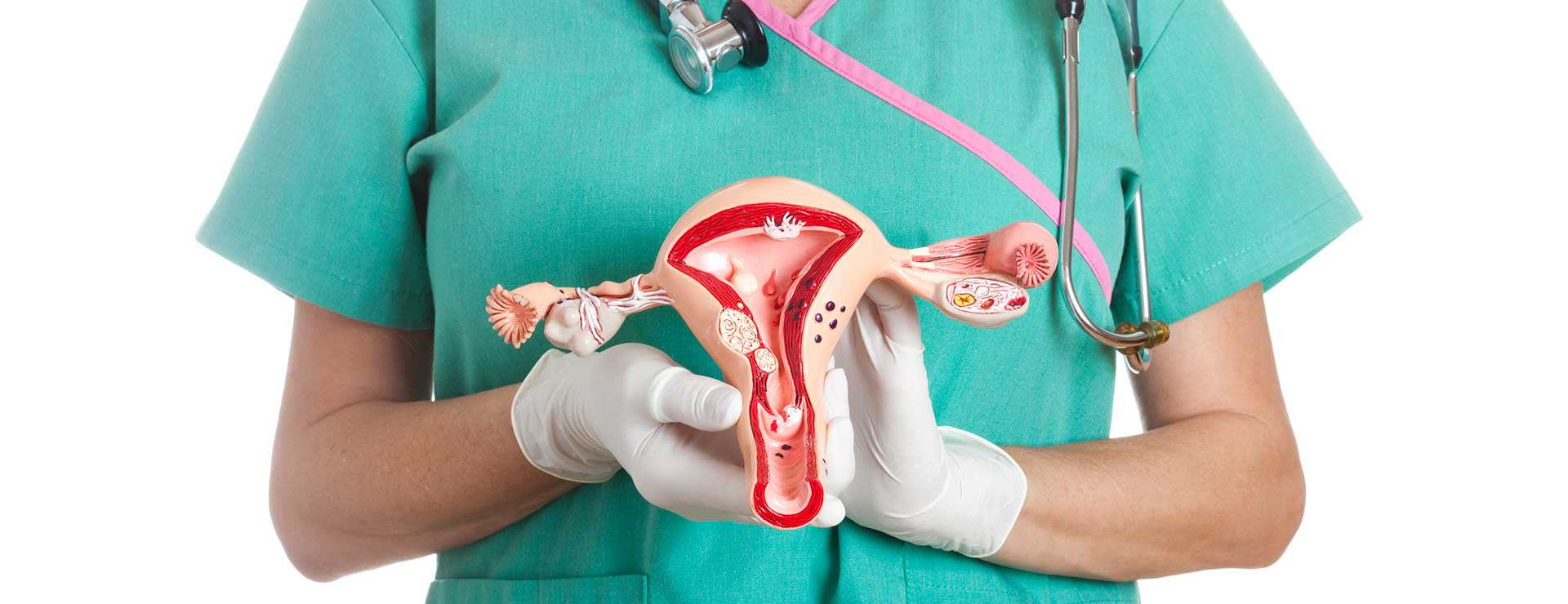
Abdominal Wall - Atlas of Anatomy from doctorlib.info The pelvis is the lower portion of the trunk, located between the abdomen and the lower limbs. • divided into the true and false pelvis by the iliopectineal line. The right and left hip bones also converge anteriorly to attach to each other. The uterus represents the essential landmark of pelvic anatomy. The pelvis (plural pelves or pelvises) is either the lower part of the trunk of the human body between the abdomen and the thighs (sometimes also called pelvic region of the trunk) or the skeleton embedded in it (sometimes also called bony pelvis, or pelvic skeleton). The lining of the uterus. It's located between the abdomen and the legs. For the purposes of the national registry exam, understanding the positioning of major bones, muscles, and ligaments is key.
It is located in the middle of the pelvis between the urinary bladder lying before and the large bowel lying behind it.
The pelvic bones are smaller and narrower. The inferior hypogastric plexus is also known as the pelvic ganglion. The pelvic floor muscles form part of the pelvic floor and play a critical role in sexual function as well as the maintenance of urinary and faecal continence, anatomy of the prostate gland edit source The nerves of the pelvis include: The pelvic girdle (hip girdle) is formed by a single bone, the hip bone or coxal bone (coxal = hip), which serves as the attachment point for each lower limb. Use the mouse scroll wheel to move the images up and down alternatively use the tiny arrows (>>) on both side of the image to move the images.>>) on both side of the image to move the images. The pelvis is a basin shaped bony structure formed by the combination of two pelvic bones (hip bones or innominate bones) and the sacrum. Gross anatomy of the pelvis—namely the bladder, uterus, fallopian tubes, ovaries, rectum, and the muscles—has remained unchanged; It provides attachment to some important muscles in the region, and forms a cavity which accommodates several important internal organs. The pelvis is the lower part of the torso. Regarding the surface anatomy, the perineal area is the region between the thighs, extending from the pubic symphysis anteriorly to the gluteal folds posteriorly. The pelvis (plural pelves or pelvises) is either the lower part of the trunk of the human body between the abdomen and the thighs (sometimes also called pelvic region of the trunk) or the skeleton embedded in it (sometimes also called bony pelvis, or pelvic skeleton). The pelvic girdle and pelvic spine.
A pelvic ultrasound is a noninvasive diagnostic exam that produces images that are used to assess organs and structures within the female pelvis. The pelvis's frame is made up of the bones of the pelvis, which connect the axial skeleton to the femurs, and therefore acts in weight bearing of the upper body. Anatomy of female pelvic area facebook twitter linkedin pinterest print fertility and reproductive health pelvic floor disorders fertility, pregnancy and childbirth women's health. Ultrasound uses a transducer that sends out. On a sagittal plane, the uterus has a pyriform shape:
Anatomy Of The Female Pelvis from www.hopkinsmedicine.org The right and left hip bones also converge anteriorly to attach to each other. However, knowledge of the anatomy of various structures that surround these organs has evolved over time. The pelvic girdle and pelvic spine. The pelvic bones are smaller and narrower. There are four articulations within the pelvis: It is located in the middle of the pelvis between the urinary bladder lying before and the large bowel lying behind it. The pelvis's frame is made up of the bones of the pelvis, which connect the axial skeleton to the femurs, and therefore acts in weight bearing of the upper body. Gross anatomy of the pelvis—namely the bladder, uterus, fallopian tubes, ovaries, rectum, and the muscles—has remained unchanged;
The pelvic region is the area between the trunk — or main body — and the lower extremities, or legs.
Pelvic anatomy www.freelivedoctor.com slideshare uses cookies to improve functionality and performance, and to provide you with relevant advertising. The uterus represents the essential landmark of pelvic anatomy. The right and left hip bones also converge anteriorly to attach to each other. The pelvis (plural pelves or pelvises) is either the lower part of the trunk of the human body between the abdomen and the thighs (sometimes also called pelvic region of the trunk) or the skeleton embedded in it (sometimes also called bony pelvis, or pelvic skeleton). It provides attachment to some important muscles in the region, and forms a cavity which accommodates several important internal organs. Classic anatomical studies have provided few details of the inferior hypogastric plexus morphology or the location and nature of the associated nerves. Cookies allow us to analyze and store information such as the characteristics of your device as well as certain personal data (e.g., ip addresses, navigation, usage or geolocation data, unique identifiers). • pelvis begins at the iliac crests and ends at the symphysis pubis. Use the mouse scroll wheel to move the images up and down alternatively use the tiny arrows (>>) on both side of the image to move the images.>>) on both side of the image to move the images. Gross anatomy of the pelvis—namely the bladder, uterus, fallopian tubes, ovaries, rectum, and the muscles—has remained unchanged; It is strengthened and supported by several joints and ligaments. The pelvis is the lower portion of the trunk, located between the abdomen and the lower limbs. The pelvis's frame is made up of the bones of the pelvis, which connect the axial skeleton to the femurs, and therefore acts in weight bearing of the upper body.
The pelvis (plural pelves or pelvises) is either the lower part of the trunk of the human body between the abdomen and the thighs (sometimes also called pelvic region of the trunk) or the skeleton embedded in it (sometimes also called bony pelvis, or pelvic skeleton). The inferior hypogastric plexus is also known as the pelvic ganglion. Each hip bone, in turn, is firmly joined to the axial skeleton via its attachment to the sacrum of the vertebral column. Classic anatomical studies have provided few details of the inferior hypogastric plexus morphology or the location and nature of the associated nerves. If you continue browsing the site, you agree to the use of cookies on this website.
3d works by Alfredo Rosales at Coroflot.com from s3images.coroflot.com Regarding the surface anatomy, the perineal area is the region between the thighs, extending from the pubic symphysis anteriorly to the gluteal folds posteriorly. The inferior hypogastric plexus is also known as the pelvic ganglion. The pelvis is the lower portion of the trunk, located between the abdomen and the lower limbs. The male pelvic floor is a complex structure made up of muscles, ligaments, nerves and fascia. Pelvic anatomy during this time of information gathering about your hysterectomy options, there are a few terms that are helpful to know about pelvic anatomy. It is inferior to the pelvic diaphragm. The uterus represents the essential landmark of pelvic anatomy. Cookies allow us to analyze and store information such as the characteristics of your device as well as certain personal data (e.g., ip addresses, navigation, usage or geolocation data, unique identifiers).
The male pelvic floor is a complex structure made up of muscles, ligaments, nerves and fascia.
The pelvis's frame is made up of the bones of the pelvis, which connect the axial skeleton to the femurs, and therefore acts in weight bearing of the upper body. By continuing to browse this site you are agreeing to our use of cookies. • divided into the true and false pelvis by the iliopectineal line. The pelvic region is the area between the trunk — or main body — and the lower extremities, or legs. Gross anatomy of the pelvis—namely the bladder, uterus, fallopian tubes, ovaries, rectum, and the muscles—has remained unchanged; It is strengthened and supported by several joints and ligaments. The pelvis is a basin shaped bony structure formed by the combination of two pelvic bones (hip bones or innominate bones) and the sacrum. It provides attachment to some important muscles in the region, and forms a cavity which accommodates several important internal organs. The pelvic girdle (hip girdle) is formed by a single bone, the hip bone or coxal bone (coxal = hip), which serves as the attachment point for each lower limb. For the purposes of the national registry exam, understanding the positioning of major bones, muscles, and ligaments is key. This mri male pelvis axial cross sectional anatomy tool is absolutely free to use. The male pelvis is different from a female's. The pelvic girdle and pelvic spine.